Hipvasc – A quantitative method to assess blood flow to the femoral head
Recently, several IPSG members travelled to Dallas to learn how to use Hipvasc, an imaging software developed for Perthes disease MRI measurements. Several centers are obtaining perfusion MRI scans of the hip in Perthes disease, which is a MRI protocol that specifically looks for how much blood flow to the femoral head is normal or abnormal. It is often difficult to visually estimate the regions of normal and abnormal blood flow to the femoral head on a perfusion MRI scan. Hipvasc is a software that uses a custom algorithm that calculates the amount of femoral head that has adequate blood flow in a perfusion MRI. An example above demonstrates a typical Hipvasc output of several slices of a hip perfusion MRI scan. In this particular hip, the more red shaded regions indicate good blood flow, while the bluer regions of the hip indicate poor blood flow. IPSG members hope to
What are the limitations with Perthes and weight bearing?
This content is restricted to subscribers
Jasta’s Advice
This content is restricted to subscribers
How do you choose between nonoperative and operative treatment for Perthes?
This content is restricted to subscribers
Dr. Sankar – CHOP
Wudbhav N. Sankar, MD Dr. Sankar is the director of the Young Adult Hip Preservation Program at Children’s Hospital of Philadelphia (CHOP). Learn more about Dr. Sankar here. Education Medical: MD – University of Pennsylvania School of Medicine, Philadelphia, PA Internship: Surgery – Hospital of the University of Pennsylvania, Philadelphia, PA Residency: Orthopedic Surgery –
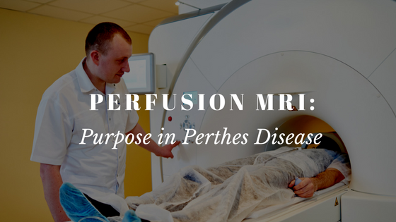
Perfusion MRIs: Recent research showing the purpose with Perthes Disease
A recent study conducted by Dr. Harry Kim, Chairman of IPSG and staff doctors and researchersat Texas Scottish Rite Hospital, looked at the use of an advancedMRI called perfusion MRI, a new diagnostic imaging testtodetermine revascularization of femoral heads of patients with Perthes disease. Revascularization is the return of blood flow to the femoral head
IPSG on Facebook
[custom-facebook-feed carousel=”true”]
Video Archives
How do you choose between nonoperative and operative treatment for Perthes?
What is containment treatment for Perthes disease?
What are the limitations with Perthes and weight bearing?
Why are there such varied treatments of Perthes among doctors?
What are current studies being carried out by IPSG?
Are there any advancements in Perthes treatment?
How frequent are flare-ups in the fragmentation stage? How do you deal with them?
What does the future of Perthes look like?
Why did you join the IPSG?
What are we learning from IPSG?
What is the International Perthes Study Group?
IPSG Website Welcome
IPSG Community Live Stream
No Results Found
The page you requested could not be found. Try refining your search, or use the navigation above to locate the post.


 Subscribe to IPSG's Channel