Perfusion MRIs: Recent research showing the purpose with Perthes Disease
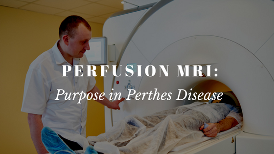
A recent study conducted by Dr. Harry Kim, Chairman of IPSG and staff doctors and researchersat Texas Scottish Rite Hospital, looked at the use of an advancedMRI called perfusion MRI, a new diagnostic imaging testtodetermine revascularization of femoral heads of patients with Perthes disease. Revascularization is the return of blood flow to the femoral head (the ballof the hipjoint) that has suffered from stoppage of blood supply. One of the most important components in the healing of Perthes is the return of blood flow to necrotic bone. Since this is such an essential part of healing, researchers wanted to better understand the things that affect the rate and quality of revascularization. The study included29 patients with an average age of 8 years who were in the first two stages of Perthes, so called Walsenstrom stage 1 or 2. The 29 patients included in this study had two or more perfusion
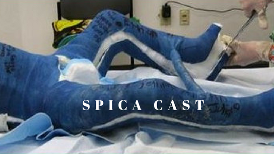
Spica Casting 101
This content is restricted to subscribers
IPSG Decides on Study Protocol for Patients who develop Perthes disease after age 11
The IPSG presented an e-Poster at the annual POSNA meeting in Toronto on the accomplishments of the study group to date. The IPSG has graciously been supported by a POSNA Clinical Trial Planning Grant and was happy to present the POSNA membership with updates on their progress. Dr Harry Kim of Texas Scottish Rite Hospital
Does contrast-Enhanced MRI more clearly depict femoral head area of necrosis than non-contrast MRI?
Current treatments for Perthes disease are most effective when applied during the early stages of Perthes disease, prior to the femoral head collapsing and breaking apart. Radiographic prognosticators during these early stages of Perthes Disease are needed to guide treatment decisions. Commonly, patients receive x-rays to evaluate the extent of involvement of the femoral head.
Can the radiographic outcomes of Legg-Calvé-Perthes be reliably assessed at skeletal maturity?
Traditionally, the outcome of treatment of Perthes disease has been assessed by characterizing the shape of the femoral head, the structure of the acetabulum, and the interface between those two hip joint structures. The most common classification system in use today is that developed by Stulberg et. al. that categorizes the affected hip into one
IPSG on Facebook
[custom-facebook-feed carousel=”true”]
Video Archives
How do you choose between nonoperative and operative treatment for Perthes?
What is containment treatment for Perthes disease?
What are the limitations with Perthes and weight bearing?
Why are there such varied treatments of Perthes among doctors?
What are current studies being carried out by IPSG?
Are there any advancements in Perthes treatment?
How frequent are flare-ups in the fragmentation stage? How do you deal with them?
What does the future of Perthes look like?
Why did you join the IPSG?
What are we learning from IPSG?
What is the International Perthes Study Group?
IPSG Website Welcome
IPSG Community Live Stream
No Results Found
The page you requested could not be found. Try refining your search, or use the navigation above to locate the post.


 Subscribe to IPSG's Channel