MRI and sonography in Legg-Calvé-Perthes disease: clinical relevance of containment and influence on treatment. Journal of Children’s Orthopaedics, 2018 October
Jandl NM, Schmidt T, Schulz M, Rüther W, Stuecker MHF
In a recent study, ultrasound measurements were correlated with magnetic resonance imaging (MRI) measurements to evaluate the utility in determing how contained the femoral head was in the acetabulum (hip socket).
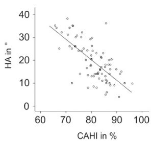
In Perthes disease, one of the goals of treatment is containment of the femoral head in the acetabulum. In children affected with Perthes disease, much of the femoral head is cartilage before it turns into bone. The difficulty in assessing the true amount of containment on xrays is that the cartilage of the femoral head is not visible on xrays. MRI clearly shows the hip cartilage, though there can be challenges with MRI including the need for sedation in young children, and cost. An advantage of ultrasound is that it is non-invasive, does not require sedation, and is inexpensive. If one is interested in evaluating how much of the femoral head cartilage is contained by the acetabulum, ultrasound may be a reasonable alternative to MRI. This study correlated ultrasound measures of the hip cartilage on patients who also had MRI of the hip, and demonstrated that ultrasound assessment of containment of the femoral head correlated (r = -0.69) well with MRI measurements of containment.

