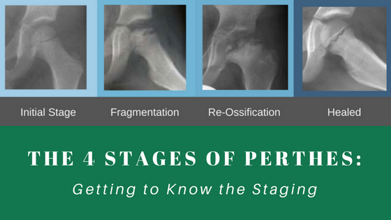
Perthes disease has four stages. The first stage, also known as the onset or necrosis stage, begins when the child first begins to limp and complain of pain. From the initial stage, the disease progresses as the body attempts to remove the damaged bone in order to begin new bone growth.
The Perthes stages include: onset stage; fragmentation stage; reconstitution stage; residual stage. Each stage of Perthes disease has key indicators, which signifies the progression of the disease through the various stages.
Onset (Synovitis, Necrosis, or Initial) Stage:
- A limp and pain begins.
- Damage occurs when blood flow to the center of the hip stops working.
- Pain is usually mild and x-rays may appear normal.
- A crescent sign (crescent shaped crack) and a break can be seen under the joint surface.
- Duration: a few weeks to months.
Fragmentation Stage
- Stage where the body attempts to repair itself, but also the stage when most of the femoral head fragmentation and collapse occurs.
- Brittle bone that lost its blood supply must be removed so that the body can grow new, healthy bone.
- On x-rays, the ball of the hip appears broken up thus the term fragmentations. It sometimes looks like Swiss cheese with patchy holes, which are the areas where the dead bone is being removed by repairing cells.
- The biggest concern at this stage is that the bone is at the greatest risk of collapsing – which can permanently deform the ball.
- The outer part of the ball may also come outside of the joint.
- Containment treatment is used in this stage if a part of the ball comes out of the joint. The ball is contained in the hip socket by casting, bracing, or surgery so that the socket can act as a mold to keep the ball round while the dead bone is adsorbed and new bone grows.
- The main goal is to keep the ball in the socket during the fragmentation stage.
- Containment is more effective if the treatment begins before the ball collapses (the ball does not collapse in every case of Perthes).
- Duration: 6 months to a year.
Reconstitution Stage (Reossification stage)
- Occurs when all the dead bone, which appears white on x-ray has dissolved.
- The shape of the ball at this stage may still improve as the patient grows. This is one of the reasons why a younger child affected with Perthes do better than an older child, since a younger child has more growth left.
- Containment treatment at this stage is no longer an effective treatment plan.
- Bone begins to fill in the holes of the ball. This is the stage when the ball starts to become bigger than the normal size in some children with severe Perthes. This is called coxa magna (big head).
- Growth of the femoral neck may also be affected and shows up as short and broad neck below the enlarged ball.
- This stage usually becomes present by 18 months after the first onset of pain or limb occurs
It could take 2 to 3 years for the entire ball to be filled with new bone. - The child can return to full activity once the round, slightly enlarged shape of the ball is becoming visible on the x-ray.
Residual Stage (Healed Stage)
- Occurs when the ball has been replaced with bone.
- When the ball is round and the shape of the upper thighbone is normal, long-term outcome is very good and no arthritis is expected in most patients.
- If the ball is oval shape, arthritis could develop by age 50 to 60 in about 50% of the patients.
- The worst residual hips are flat or saddle shaped – the chance of getting early arthritis is higher.
- This is one of the reasons why we treat a child with Perthes early in order to prevent the ball from becoming very flat.

